Radiography
|
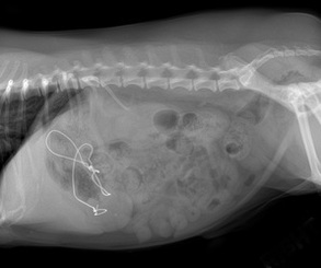
Central Vet Imaging Radiologists interpret a variety of x-ray studies daily. These studies include both small and large animal imaging.
Radiology is the most common imaging modality in Veterinary Medicine and is a common source of referrals. Our experienced board certified Radiologists have an incredible eye for the smallest details that can be found in these studies, which can improve case management and outcome. They possess the medical knowledge to prioritize radiographic findings and convert them into a diagnosis that can guide successful treatment. Our Radiologists assist primary and specialty veterinarians in getting as much information as possible from their radiographic studies, facilitating quicker more accurate diagnosis and guiding treatment as directly as possible. Radiographic interpretation by a board certified Radiologist is not only for ill patients; geriatric screening, wellness and pre-anesthetic exams, pre-breeding exams and pregnancy evaluation are also performed on a routine basis. Radiographs are normally sent directly from the referring veterinarian by a web-based method called TELEMEDICINE. If your veterinarian does not have telemedicine capability or radiography has not be performed yet, please CONTACT US to help create a plan for your pet. |
Ultrasound Services |
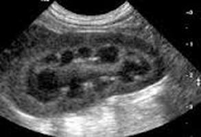
We work closely with your primary veterinarian to perform outpatient ultrasound services. Dr. Jones can perform ultrasound of almost any body area to help diagnose or rule out a variety of disorders that can affect pets. He routinely performs examinations of the abdomen, heart, neck, thorax, or even mass lesions on a limb. Typical organs we look at during our abdominal ultrasound studies includes: liver, spleen, kidneys, adrenal glands, bladder, stomach, intestines and pancreas. Ultrasound of the chest is commonly performed to look for causes of abnormal fluid accumulation, evaluate mass lesions such as lung tumors or abscesses, and assess heart disease. Abnormalities found with an ultrasound exam can be sampled for definitive diagnosis by performing an ultrasound guided biopsy or fine needle aspirate (FNA) at the time of the scan for microscopic evaluation.
(Want additional information on when to use ultrasound? Click here) |
Echocardiography
|
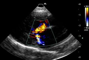
Echocardiography is the study of heart anatomy and function via the use of ultrasound imaging. Dr. Jones routinely performs evaluation of the heart of dogs, cats, and other animals to evaluate the cause of heart murmurs, arrthythmias, or radiographic abnormalities. Through use of this imaging modality, we are able to diagnose heart problems such as valve disease, congenital heart defects, congestive heart failure, cardiomyopathy, endocarditis, and pulmonary hypertension. Echocardiography allows us to better determine the cause of symptoms seen at home (such as coughing or exercise intolerance). We work closely with your pet's primary veterinarian to elucidate the cause of your pet's illness and an echo study is an excellent way of ruling in or ruling out primary heart disease. We also routinely perform echo studies as a screening tool for breeders and pet owners who have breeds known to be predisposed to a variety of heart problems (Maine Coons, Boxers, Dobermans, Persians, Cavalier King Charles Spaniels, and others)
|
Veterinary CT Study
|
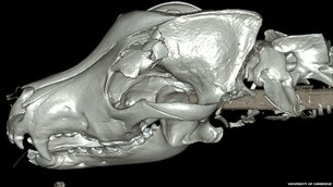
Computerized tomography — also known as CT Scan — is an innovative way of imaging both small animal and large animal patients. CVI radiologists routinely interpet CT scans to help local (and distant) veterinarians diagnose a variety of diseases. This imaging modality can be used to scan the chest for cancer, evaluate bones for tiny fractures or destructive changes not seen on conventional x-rays, outline tumor margins for surgical planning, and much more. Areas of the body that cannot be visualized adequately with ultrasound are better seen with a CT scan.
Please note that Central Vet Imaging does not have CT scanning capability on site at our facility. Our staff only offers interpretation of studies performed at veterinary specialty and imaging centers around the United States. |
Veterinary MRI
|
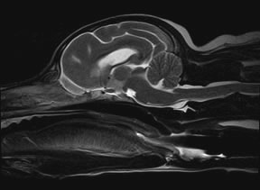
As part of our services to primary care and specialist veterinarians, our team of highly trained radiologists routinely interprets MRI studies performed on animals. MRI is a highly sophisticated and technical imaging modality used to diagnose a range of diseases, most commonly involving the nervous system, such as brain tumors or spinal disc disease. In additional to brain and spine imaging, MRI is also useful to evaluate musculoskeletal disease involving any body area. It is also quite useful for imaging areas that cannot be assessed with ultrasound, such as the pelvic canal or deep tissues of the neck. Please note that Central Vet Imaging does not have MRI scanning capability on site at our facility. Our staff only offers interpretation & read out reports of MRI studies performed at veterinary specialty and imaging centers around the United States. |

